PORPHYRINS
Porphyrins are tetrapyrrole macrocycles with conjugated double bonds and various groups attached to the perimeter. The porphyrins can accept two hydrogen ions to form +2 diacids or donate two protons to form – 2 dianions.
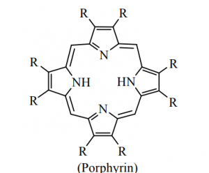
variation of substituent of facilitates the tuning of electron donating and electron withdrawing ability of the ligand.
The porphyrin and corrin ring systems are of great biological importance. Four pyrrole units are linked by – CH = bridges as shown in figure but in corrin ring one – CH = group is less.

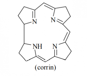
The corrin ring has 19 carbons, whereas porphyrins have 20. Also, the pyrrol like rings in corrin are fully saturated “edge-carbon” centre whereas porphyrins are highly conjugated. Because of the high number of sp3 carbon centre, corrins are more flexible than porphyrins and are not as flat. Porphyrins is aromatic in nature.
These rings are intensely colored.
Most importance of these rings in the bio-system can be illustrated as:
- Iron complex of the substituted porphin is Heme.
- When magnesium lies at the centre of substituted porphin ring; the resulting complex is calledchlorophyll.
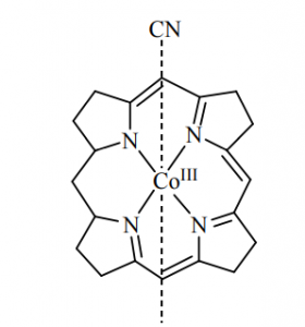
- If cobalt is the central metal atom of substituted corrin ring system, is called vitamin B12 (coblamine) Cobalt is present in +3 oxidation state in vitamin B12. Vitamin B12 is the well known naturally occurring organometallic compound. It is only vitamin known that contain metal. It may be reduced by one electron (vitamin B12 r) or two electron (vitamin B12 s) to form cobalt (II) and cobalt (I) oxidation state.
Vitamin B12 is the only known essential bio-molecule with a stable metal carbon bond.
[Vit. B Co(I)] + CH3I [Vit B12 Co(III)CH3]+ I–
Characterization of porphyrins:
- UV-visible spectroscopy: The free base porphyrin usually have four absorption bands in the range of 500 to 650 nm. In addition to the four bands, there is a band in the range of 400 to 450 nm.
This known as the “soret band” and is a characteristic of all conjugated tetrapyrroles.
- IR-spectroscopy: The NH stretching frequency band appears at 3300 cm–1.
- 1H-NMR spectroscopy: In the 1H – NMR spectroscopy the N-H proton appears at high field due to anisotropic effect. It is generally appears at – 2 to – 3 ppm.
The metalloporphyrin have no N – H proton signal appear in the NMR spectroscopy.
The UV-visible spectroscopy as one of the best too for the characterisation of porphyrin.
Porphyrins are found in many metalloenzyme:
| Enzyme | Function |
Fe-porphyrin | Cytochrome | Electron transfer |
Fe-porphyrin | Hemoglobin, Myoglobin | Dioxygen carrier |
Mg-porphyrin | Chlorophyll | Photosynthesis |
Oxygen carrier and storage devices:
Dioxygen (O2) is biologically very important. The most common mode of reaction of molecular oxygen with transition metal complexes oxidation. The reaction of dioxygen with a complex oxidation. The reaction of dioxygen with a complex so as to incorporate the dioxygen ligand with undergoing any reduction on oxygen in called oxygenation. This is contrast to oxidation reaction in which O2 looses its identity during the reaction.
Some transition metal complexes act as reversible carriers of molecular O2 i.e., they take up and release O2 reversibnly as follows:
To understand about oxygen carriers we consider electronic structure of O2 molecule.
Iron Storage:
Protein | Ions | Proteins | Ions |
Serum albumin | Cu+2, Zn2+ | Ferritin | Fe3+ |
Phosphoprotein | Ca+2 | Metallothionein Ceruloplasmin | Cu2+, Zn2+ Cu2+, Zn2+ |
Transport and storage of molecular oxygen asian essential physiological function.
Example | Metal | Mole ratio (O2/metal) | Function | Ligands |
Hemoglobin (Hb) | Fe(II) | 1/1 | Carrier | Porphyrin |
Myoglobin (Mb) | Fe(II) | 1/1 | Storage | Porphyrin |
Hemerythrin | Fe(II) | 1/2 | Storage | Protein |
Hemocyanin | Cu(I) | 1/2 | Carrier | Protein |
Iron containing oxygen carrier are present inside the cells and copper containing oxygen carrier are found in extracellular fluids.
Both heme and non-heme iron protein are involved in oxygen transport and storage. Heme O2 carrier are responsible for red colour of human blood, while the blue pigment of crab blood, hemocyanin. Some marine worms have a violet colour, which is due to presence of non-heme iron-protein hemerythrin.
Hemoglobin (Hb) and Myoglobin (Mb) in oxygen transport mechanism:
The best known iron porphyrin compounds are haemoglobin and myoglobin, oxygen transfer and storage agents in the blood and muscle tissue, respectively. The human body contains about 4g of iron. About 70% of this is found as haemoglobin, the red pigment in the erythrocytes (red blood cells). Most of the rest is stored as ferretin.
Hemoglobin occurs in all vertebrates (with certain exception) and in many invertebrates, it has also been found in certain strains of yeasts, mould etc.
Fe-porphyrin referred to as heme is the prosthetic group of hemoglobin (Hb) and Myoglobin (Mb). Each Hemoglobin (Hb) molecule has four heme groups bound to the globin on its surface. On each heme unit of Hemoglobin (Hb) the four square-planar coordination sites of the Fe(II) are occupied by porphyrin nitrogen atom. Whereas the fifth coordination site of iron occupied by the nitrogen of histidine (distal histidine) ligand of the protein chain i.e. globin chain, and the sixth position of iron remains vacant occupied by H2O in their deoxy forms and this site is occupied by O2 in their oxy forms. The distal histidine protects Hb and Mb-form CO poisoning. Myoglobin (Mb) is a monomer having only one heme unit. Hemoglobin and myoglobin are transport and storage of dioxygen respectively.
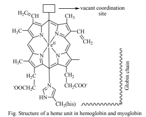
The planar porphyrin ring of heme unit of Hb and Mb, due to the presence of conjugated double bond in the porphyrin, stable
and low lying
orbital are available and these allow the characteristic charge transfer electronic transition to give the red colour of blood.
Function of Hb and Mb:
Hemoglobin (Hb carries O2 from lungs to tissues where it is transfer to myoglobin (Mb) and stored there in for metabolic requirements. To make this process thermodynamic possible, the oxygen affinity of Hbn in lungs where oxygen concentration is high should greater than of Mb and reverse condition should arise in the tissue where oxygen concentration is less.
Figure show that Hb is about as good on O2 binder as Mb at high O2 pressure. It is much poorer at the lower pressures prevailing in muscle and hence passes its oxygen on to Mb as required. Moreover, the need for O2 will be greatest in tissues where O2 is consumed followed by production of CO2. The CO2 lowers the pH, thus causing the Hb to release even more oxygen to Mb. The pH sensitivity (Bohr effect) as well as the progressive increase of O2 binding constants in Hb are due to the interaction between the subunits, Mb behaves more simple because it consists of only one unit. Thus, each of the two is essential in the complete oxygen transport process.
Oxygenation equilibria of haemoglobin (Hb) and myoglobin (Mb)
Cooperative Effect
Coordination of one O2 leads to conformational changes in the protein chain leading to facilitated coordination of O2 by the other 3 sub-units in hemoglobin.
Both Hb and Mb bind with oxygen but first one act as carrier and later one as storage device in human body. It raises interesting point why one act as carrier and other as storage devices while both have same heme unit. The difference lies on structure of both Hb and Mb and on the environment of the human body where these have to show their function. Hb consists of four unit of heme group while Mb has single heme unit. Heme unit is embedded in hydrophobic proteins environment (Globin proteins) in both biological devices. The hydrophobic protein environment protects the oxidation of Fe+2 to Fe+3. In case of Hb system, four heme units cooperate to each other and adjust their structure according to their comfort. This behaviour is probably due presence of -helix of proteins in which act like a spring and connecting to these heme units. In Hb as the one O2 binds, the molecular shape change such a way that those additional O2 molecules could bind. This is a kind of communication among fourth heme units of Hb makes O2 binding process very easier. It is interesting that the four equilibrium constants (K1, K2, K3, K4) respective to four step reaction equilibria become in increasing order (K1 < K2 < K3 < K4) while one expect in reverse order. Not only this, even K1 (5 to 60) is much smaller than K4 (3000 to 6000).
In similar way as one O2 get removed from Hb, it triggers the release of remainder O2 molecules. This whole phenomenon is called as “cooperative effect.” The removal of oxygen is also favoured by pH change. When CO2 concentration in capillaries is enhanced, it supports the following reaction in forward direction which increase the acidity.
This decreased in pH favoures the release of O2 from Hb. This effect is called as Bohr Effect. These are two important factors which make labile binding of O2 and easier removal. In the Hb and supports the carrier activity of Hb.
Derivation of Hill Equation for Oxygenation
Myoglobin has one heme group per molecles and serves as storage in muscles but binding mechanism of O2 is similar to Hb.
This equation is known as Hill equation and the exponent n is called the Hill constant.
The data obtained from Hb between 10 and 90% oxygenation can be fitted to the Hill equation to give vary of n ~ 3 for normal Hb. The form of oxygenation curve and that the fact if fits n > 1 indicated that the cooperative interaction between the subunits. The addition of oxygen to a subunit affects the oxygen affinity other subunits, this is an example of Allosteric effect (Entatic effect) literally means “a stretched state or state being under tension”. n < 4 indicates that the cooperative interaction between heme units is rather moderate.
The cooperative interaction on where the binding of one molecule of a substance influences the binding of molecule of the same kind is described to as a homotropic allosteric interaction.
Mechanism of homotropic allosteric effect of O2 in Hb:
On oxygenation, a trigger mechanism proposed by perutz operates through the heme-heme interaction to carry out the change. T-form to R-form. To this change, researcher suggested that in deoxy-Hb, Fe(II) remains in high spin state, but on oxygen Fe(II) attains the low spin state. This spin state change acts as the trigger.
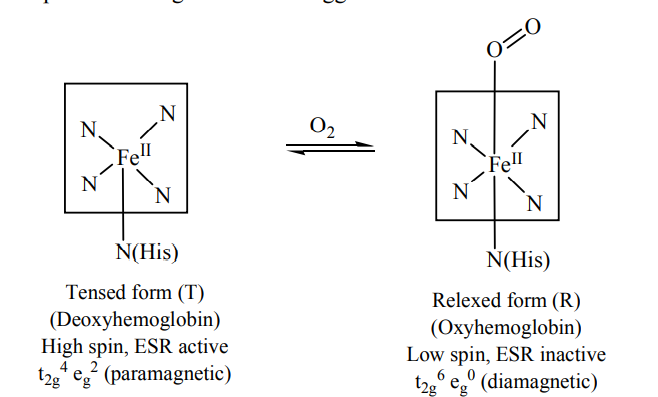
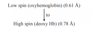
The Fe(II) – N bond length (high spin Fe(II) and sp2N) is ~218 pm. The eg electrons directly interact with the ligands and thus the removal of eg electrons in attaining the low spin stat reduces the bond length. The size of porphyrin cavity allows the sitting of the metal having the M – N bond length ~ 200-205 pm. Thus the high spin Fe(II) is deoxy Hb cannot sit in the porphyrin cavity. In fact, the size of Fe2+ increase by 28% on going from
The globular protein prevents the irreversible oxidation of Fe(II) to Fe(III).
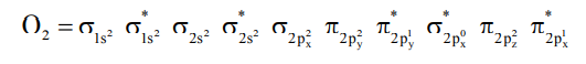
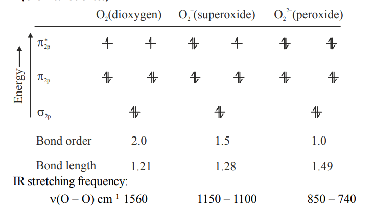
Here, it should be pointed out that there are evidences to support that the oxygenated form of Hb, iron exists as Fe(III) and O2 as O2– (superoxide) because of the, O – O stretching frequency ~ 1106 cm–1 is close to that of O– (
– O = 1097 cm–1). The Fe – O – O bond angle is close to 120º. In fact, Fe (II) is reversible oxidised to Fe(III) in this O2– uptake process and at the site of delivery of O2, it again attains the Fe(II) state through deoxygenation.
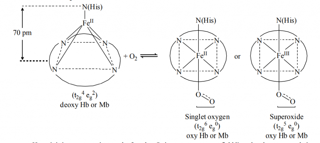
Here, it is important point out the fact that O2 is not a very strong field ligand and consequently it is not expected to carry out the desired spin pairing on oxygenation of Hb. It is believed that the heme unit is properly tuned by its substituents to make the spin pairing easier. Thus, the oxy forms of Hb and Mb, O2 remains as a singlet form. The singlet O2 is a good -acceptor and it can act as a fairly strong field ligand to induce the spin pairing in Fe(II).
Importance of glubin (protein chain):
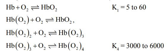
Allosteric effect of O2, H+, CO2, Cl– and diphosphoglycerate (DPG) on O2 affinity of Hb.
Protection of Hb and Mb from irreversible oxidation by O2
Weakening the interaction of CO with heme and stabilizing the binding of O2 by histidine reversible.
– donor properties of the imidazole moitey of the proximal histidine stabilise the O2 binding by the
– acceptance property of O2.
Protonation and deprotonation sites in globin protein chain are important in maintaining the biological pH and CO2 transport.
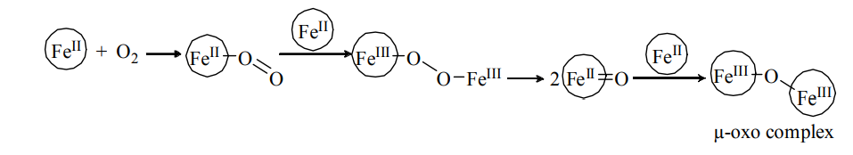
Prevent the irreversible µ – oxo complex formation.
Some synthetic complexes as such CO (acacen) complex and triphenylphosphine) iridium known as Vaska’s Iridium complex show direct oxygenation and help to understand oxygen binding in biological system.
Poisoning towards hemoglobin and myoglobin:
Different
-acid ligands like CO, NO, PF3 etc. which are electrically neutral and not much bulky can competitively replace O2 from the sixth octahedral site of Hb and Mb. Consequently, the O2 transport mechanism gets arrested and toxicity arieses. CN– may also bind the site, but the heme pocket surrounded by the hydrophobic environment does not welcome CN– much. The
– bonding ligands like NH3 and amines may also block the oxygenation site.
The CN– actually blocks the cytochrome C oxidase involved in the respiratory chain. To remove the bound CN– from cytochrome C oxidase, some methe-Hb (oxy-hemoglobin) are to be generated either by inhalation of amylnitrite vapour or by injection of NaNO2 solution. Met-Hb bearing Fe can bind CN– more strongly than the cytochrome C oxidase. Consequently. CN– removed from the respiratory chain to regenerate the electron tunneling path.


